E-ISSN:2320-8686
P-ISSN:2321-127X
RNI:MPENG/2017/74037

Case Report

Carotid Artery

International Journal of Medical Research and Review

2025 Volume 13 Number 2 Apr-Jun
Publisherwww.medresearch.in

Unmasking The Culprit – Pseudoaneurysm of Petrous Internal Carotid Artery

Anju KR1*, Swapna UP2, Nikhila R3, Anjali V4
DOI:https://doi.org/10.17511/ijmrr.2025.i02.07

1* Anju Krishna R, Junior Resident, Department of ENT, Sree Uthradom Thirunal Academy of Medical Sciences, Vattappara, Trivandrum, Kerala, India.

2 Swapna UP, Professor and HOD, Department of ENT, Sree Uthradom Thirunal Academy of Medical Sciences, Vattappara, Trivandrum, Kerala, India.

3 Nikhila Rajendran, Assistant Professor, Department of ENT, Sree Uthradom Thirunal Academy of Medical Sciences, Vattappara, Trivandrum, Kerala, India.

4 Anjali Vinod, Assistant Professor, Department of ENT, Sree Uthradom Thirunal Academy of Medical Sciences, Vattappara, Trivandrum, Kerala, India.

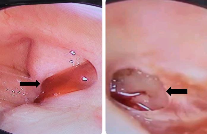
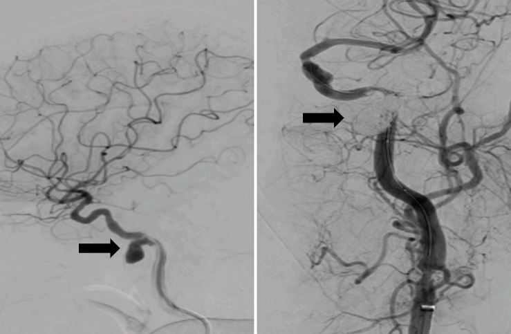
Pseudoaneurysms of the petrous internal carotid artery are rare. This is the case of a patient who presented with intermittent episodes of profuse bleeding from the left nostril and pulsatile tinnitus. Digital subtraction angiography (DSA) showed a pseudoaneurysm from the left petrous internal carotid artery which was promptly treated with balloon-assisted coiling.

Keywords: Pseudoaneurysm, epistaxis, internal carotid artery, balloon occlusion test, endovascular coiling

Corresponding Author How to Cite this Article To Browse
Anju Krishna R, Junior Resident, Department of ENT, Sree Uthradom Thirunal Academy of Medical Sciences, Vattappara, Trivandrum, Kerala, India.
Email:
Anju KR, Swapna UP, Nikhila R, Anjali V, Unmasking The Culprit – Pseudoaneurysm of Petrous Internal Carotid Artery. Int J Med Res Rev. 2025;13(2):34-36.
Available From
https://ijmrr.medresearch.in/index.php/ijmrr/article/view/1545

Manuscript Received Review Round 1 Review Round 2 Review Round 3 Accepted
2025-03-08 2025-03-18 2025-03-28 2025-04-08 2025-04-18
Conflict of Interest Funding Ethical Approval Plagiarism X-checker Note
None Nill Yes 11.36

© 2025 by Anju KR, Swapna UP, Nikhila R, Anjali V and Published by Siddharth Health Research and Social Welfare Society. This is an Open Access article licensed under a Creative Commons Attribution 4.0 International License https://creativecommons.org/licenses/by/4.0/ unported [CC BY 4.0].

Download PDFBack To ArticleIntroductionCase ReportDiscussionConclusionReferences

Introduction

Pseudoaneurysms of the petrous internal carotid artery are rare and result from head trauma, radiation therapy [1], surgical complications, tumour invasion, fibromuscular disease and chronic otitis media [2], [3].

Most cases are discovered incidentally on computed tomography (CT) scans. Management depends on the clinical presentation of the patient and the various options include close observation, trapping with endovascular balloon occlusion, proximal occlusion, coil embolization or stent placement with or without coil embolization, surgical trapping and revascularization through a high-flow bypass [4]

Case Report

A 69-year-old female with a history of hypertension, coronary artery disease and recurrent episodes of left ear discharge since childhood presented with intermittent episodes of profuse bleeding from the left nostril associated with pulsatile tinnitus of two weeks duration. Nasal endoscopy revealed bleeding through the left eustachian tube (ET) orifice and otoendoscopy of the left ear showed bloodstained pulsatile discharge from medium central perforation (Fig 1).

Bleeding and coagulation parameters were normal. Computed tomography of the nose and paranasal sinus was normal. HRCT Temporal bone showed a vascular lesion arising from the left internal carotid artery (ICA). Digital Subtraction angiography revealed a pseudoaneurysm of the left petrous internal carotid artery with severe narrowing of the lateral segment of ICA distal to aneurysm (Fig 2).

ijmrr-1545-01.JPG
Figure 1:
Nasal endoscopy and Otoendoscopy of left ear.

ijmrr-1545-02.JPG
Figure 2:
DSA pre and post-endovascular coiling 

A balloon occlusion test was done to assess the contralateral blood flow before parent artery occlusion and was found to be normal. The patient underwent near complete coiling of the left petrous internal carotid artery (Fig 2) achieving a residual filling of class 2 according to Modified Raymond Roy classification10. Post-procedure Doppler was normal.

Discussion

Cervical ICA becomes the petrous segment as it enters the petrous temporal bone at the base of the skull anterior to the internal jugular vein and medial to the styloid process. Aneurysms arising from the petrous segment of ICA are rare. It can be true or pseudoaneurysm. Pseudoaneurysm occurs as a result of blood leakage from the injured site and is contained in a fibrous tissue capsule [5]. Such an injury occurs as a result of blunt or penetrating trauma, inflammatory reactions or infections. Infectious causes can be otomastoiditis, pharyngeal and tonsillar infections and cholesteatomas. Some cases can be congenital in origin.

It is usually asymptomatic and is diagnosed incidentally on imaging. Other presentations can be ear bleed, epistaxis, pulsatile tinnitus, facial numbness, dizziness, hearing disturbances or cranial nerve disorders [6]. CT Angiography or Magnetic Resonance Angiography are common imaging modalities, whereas DSA forms the gold standard [7].

The primary goal of treatment is to provide symptomatic relief and to minimize the risk of hemodynamic compromise [8]. Since they are secured by petrous bone, open surgical techniques are challenging and endovascular techniques are more advantageous.


Endovascular surgical approaches include balloon embolization, stand-alone stenting, coil embolization or stent-assisted coiling [9], [10]. The outcome of treatment depends on the anatomy of the segment involved, collateral flow to the brain and age of the patient.

Conclusion

Diagnosis and treatment of pseudoaneurysm of the petrous internal carotid artery is challenging. The proximity between the petrous internal carotid artery and the eustachian tube should be kept in mind for early diagnosis and prompt management in patients presenting with epistaxis.

References

1. K. M. Cheng, C. M. Chan, Y.L. Cheung, H.M. Chiu, K.W. Tang, C.K. Law. Endovascular treatment of radiation-induced petrous internal carotid artery aneurysm presenting with acute haemorrhage. A report of two cases. Acta Neurochir (Wien), 143 (4) (2001), pp. 351-356 [Crossref][PubMed][Google Scholar]

2. M. Johnson, V. Madhavakurup, A. V. Sandeep. Pseudoaneurysm of petrous internal carotid artery presenting as aural polyp. Indian J Otol, 19 (1) (2013), pp. 27-29 [Crossref][PubMed][Google Scholar]

3. Hiranandani LH, Chandra O, Malpani NK. et al: An internal carotid aneurysm in the petrous temporal bone. J Laryngol Otol 76:703–706. 1962. [Crossref][PubMed][Google Scholar]

4. Bilateral Petrous Internal Carotid Artery Pseudoaneurysms Presenting with Sensorineural Hearing Loss Catherine J Hwang a, Gul Moonis b. AJNR Am J Neuroradiol. 2003;24(6):1139–41. . [Crossref][PubMed][Google Scholar]

5. Choi SH, Park H, Yang TK, Song CI. Pseudoaneurysm of the Petrosal Internal Carotid Artery in the Middle Ear as a Complication of Middle Ear Cholesteatoma. Journal of Audiology and Otology. 2015 Apr 20;19(1):58–61. [Crossref][PubMed][Google Scholar]

6. Chavan R, Nasli Ichaporia, Sanjay Vhora, Kaustubh Aurangabadkar, Dhanesh Kamerkar, Suhas Hardas, et al. Endovascular management of internal carotid artery pseudoaneurysms: Retrospective observational study. Interdisciplinary neurosurgery. 2021 Jun 1;24:101042–2. [Crossref][PubMed][Google Scholar]

7. Hwang CJ, Moonis G, Hurst RW, Hockstein N, Bigelow D. Bilateral petrous internal carotid artery pseudoaneurysms presenting with sensorineural hearing loss. AJNR Am J Neuroradiol. 2003 Jun-Jul;24(6):1139-41. PMID: 12812941; PMCID: PMC8149025 [Crossref][PubMed][Google Scholar]

8. Auyeung KM, Lui WM, Chow LC, Chan FL. Massive epistaxis related to petrous carotid artery pseudoaneurysm after radiation therapy: emergency treatment with covered stent in two cases. AJNR Am J Neuroradiol. 2003;24:1449–1452. [Crossref][PubMed][Google Scholar]

9. . Willinsky R, Lasjaunias P, Pruvost P, Boucherat M. Petrous internal carotid aneurysm causing epistaxis: balloon embolization with preservation of the parent vessel. Neuroradiology. 1987;29:570–572. doi: 10.1007/BF00350444 [Crossref][PubMed][Google Scholar]

10. Mascitelli J, Moyle H, Oermann E et al. An Update to the Raymond-Roy Occlusion Classification of Intracranial Aneurysms Treated with Coil Embolization. J Neurointerv Surg. 2015;7(7):496-502. doi:10.1136/neurintsurg-2014-011258 [Crossref][PubMed][Google Scholar]

Disclaimer / Publisher's NoteThe statements, opinions and data contained in all publications are solely those of the individual author(s) and contributor(s) and not of Journals and/or the editor(s). Journals and/or the editor(s) disclaim responsibility for any injury to people or property resulting from any ideas, methods, instructions or products referred to in the content.