E-ISSN:2320-8686
P-ISSN:2321-127X
RNI:MPENG/2017/74037

Case Report

Peutz-Jeghers syndrome

International Journal of Medical Research and Review

2025 Volume 13 Number 2 Apr-Jun
Publisherwww.medresearch.in

Small bowel obstruction in Peutz-Jeghers syndrome: case report

Abdelhak E1, Khalid J2, Mosaab E3*, Khadija K4, Abdessamad M5, Mounir B6, Abdelilah E7, Khalid E8, Khaleq K9
DOI:https://doi.org/10.17511/ijmrr.2025.i02.10

1 Ettaoussi Abdelhak, Medicine and Pharmacy of Casablanca, Visceral Emergency Department P35, IBN ROCHD University hospital of Casablanca, Casablanca, Morocco.

2 Jamaleddine Khalid, Medicine and Pharmacy of Casablanca, Visceral Emergency Department P35, IBN ROCHD University hospital of Casablanca, Casablanca, Morocco.

3* Elhaddadi Mosaab, Medicine and Pharmacy of Casablanca, Visceral Emergency Department P35, IBN ROCHD University hospital of Casablanca, Casablanca, Morocco.

4 Kamal Khadija, Medicine and Pharmacy of Casablanca, Visceral Emergency Department P35, IBN ROCHD University hospital of Casablanca, Casablanca, Morocco.

5 Majd Abdessamad, Medicine and Pharmacy of Casablanca, Visceral Emergency Department P35, IBN ROCHD University hospital of Casablanca, Casablanca, Morocco.

6 Bouali Mounir, Medicine and Pharmacy of Casablanca, Visceral Emergency Department P35, IBN ROCHD University hospital of Casablanca, Casablanca, Morocco.

7 Elbakouri Abdelilah, Medicine and Pharmacy of Casablanca, Visceral Emergency Department P35, IBN ROCHD University hospital of Casablanca, Casablanca, Morocco.

8 Elhattabi Khalid, Medicine and Pharmacy of Casablanca, Visceral Emergency Department P35, IBN ROCHD University hospital of Casablanca, Casablanca, Morocco.

9 Khalid Khaleq, Medicine and Pharmacy of Casablanca, Visceral Emergency Department P35, IBN ROCHD University hospital of Casablanca, Casablanca, Morocco.

Introduction: Peutz-Jeghers syndrome (PJS) is a rare autosomal dominant congenital disorder characterised by the presence of hamartomatous polyps in the gastrointestinal tract and mucocutaneous lentiginosis. It is associated with an elevated risk of cancer and substantial morbidity related to polyps, notably intestinal intussusception during childhood.

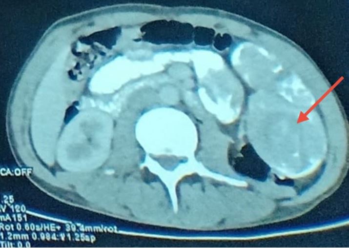
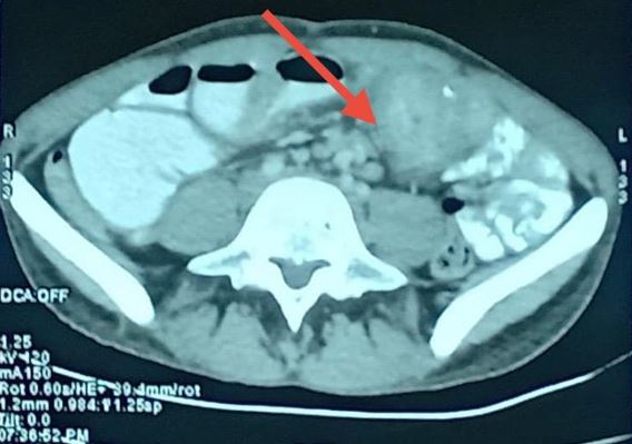
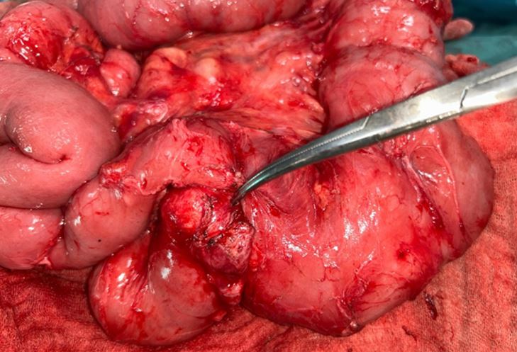
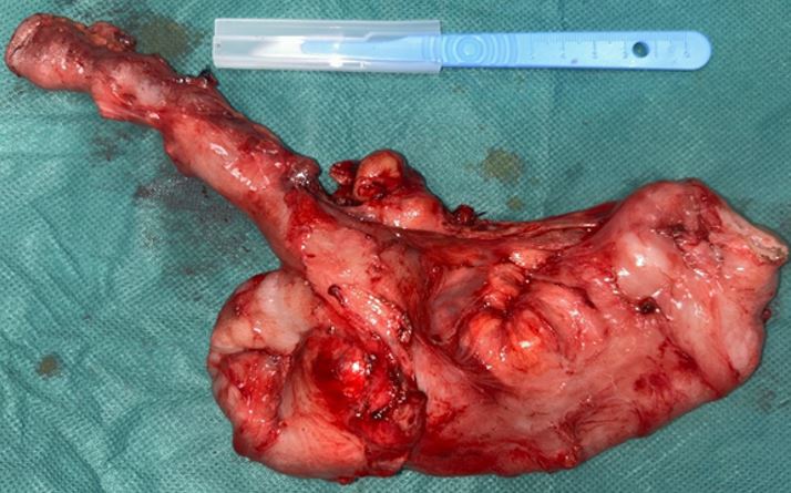
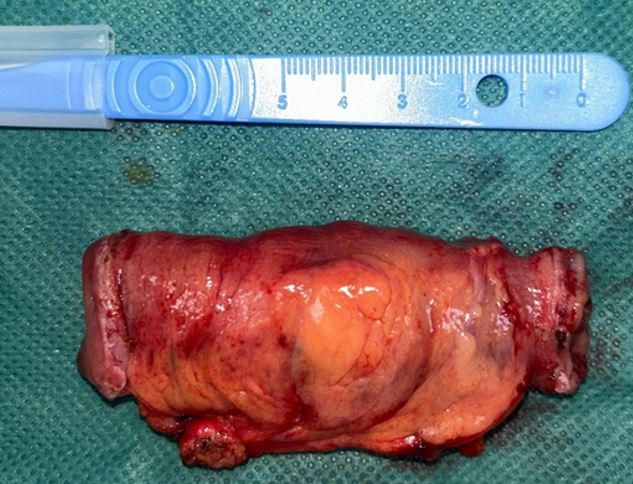
Case report: We report the case of a 32-year-old man patient, who consulted for subocclusif syndrome with multiple pigmented spots on the face and lips. Abdominal computed tomography (CT) revealed an image of jejuno-jejunal intussusception and substenotic jejunal mass. The patient underwent a double-segmental small bowel resection, removing the intussusception and the jejunal mass. The pathologic examination confirmed the diagnosis of Peutz-Jeghers polyps with moderately differentiated, invasive adenocarcinoma.

Discussion: The diagnosis of SPJ can be established in patients presenting one or more polyps and at least two of the associated clinical criteria: labial melanin deposits, family history of the syndrome and polyposis of the small bowel. Half of the cases present with small bowel obstruction. PJS is associated with an increased risk of gastrointestinal and non-gastrointestinal malignancies. Endoscopic or surgical polypectomy remains the preferred treatment option to prevent complications.

Conclusion: Regular surveillance of the gastrointestinal tract is recommended both for cancer prevention and early detection, and to prevent polyp-related complications, and certainly improve prognosis in these patients.

Keywords: Peutz-Jeghers syndrome, Hamartomatous polyp, Intussusception, Small bowel obstruction

Corresponding Author How to Cite this Article To Browse
Elhaddadi Mosaab, Medicine and Pharmacy of Casablanca, Visceral Emergency Department P35, IBN ROCHD University hospital of Casablanca, Casablanca, , Morocco.
Email:
Abdelhak E, Khalid J, Mosaab E, Khadija K, Abdessamad M, Mounir B, Abdelilah E, Khalid E, Khaleq K, Small bowel obstruction in Peutz-Jeghers syndrome: case report. Int J Med Res Rev. 2025;13(2):46-51.
Available From
https://ijmrr.medresearch.in/index.php/ijmrr/article/view/1551

Manuscript Received Review Round 1 Review Round 2 Review Round 3 Accepted
2025-05-20 2025-05-26 2025-06-03 2025-06-11 2025-06-19
Conflict of Interest Funding Ethical Approval Plagiarism X-checker Note
Authors have declared that no competing interests exist. None As per international standard or university standard, written ethical approval has been collected and preserved by the author(s). 11.32

© 2025 by Abdelhak E, Khalid J, Mosaab E, Khadija K, Abdessamad M, Mounir B, Abdelilah E, Khalid E, Khaleq K and Published by Siddharth Health Research and Social Welfare Society. This is an Open Access article licensed under a Creative Commons Attribution 4.0 International License https://creativecommons.org/licenses/by/4.0/ unported [CC BY 4.0].

Download PDFBack To ArticleIntroductionCase PresentationDiscussionConclusionReferences

Introduction

Peutz-Jeghers syndrome (PJS) is a rare autosomal dominant disorder characterised by gastrointestinal (GI) hamartomatous polyps and distinctive mucocutaneous pigmented spots, due to mutation of the STK11/LKB1 gene located on chromosome 19p. Approximately half of the cases are inherited, while the remaining are sporadic as a result of spontaneous mutation [1], [2].

Chronic anemia, intestinal occlusion by intussusception, and sporadic abdominal pain are symptoms of digestive hamartomatous polyps. The diagnosis is evoked by radiological imaging showing intussusception of the polyps. However, only a pathological examination can offer a definitive diagnosis [3].

We hereby report an uncommon case of a man experiencing intestinal intussusception due to Peutz-Jeghers syndrome, this case underscores the importance of including this syndrome in the differential diagnosis of intestinal obstructions, particularly those that are challenging to diagnose clinically, and contributes to laying the groundwork for future research on genetic predispositions, familial implications, and advances in diagnostic techniques. This case report aims to present a clinical case of intestinal intussusception in a patient with Peutz-Jeghers Syndrome (PJS), emphasising the diagnostic process, surgical management, and clinical outcome. Through this case, we aim to underscore the significance of prompt recognition of gastrointestinal complications in PJS and to contribute to the existing literature by highlighting key considerations in the acute management of intussusception in this patient population.

Case Presentation

This is the case of a 32-year-old patient who underwent surgery for appendicular peritonitis in 2003 via a midline laparotomy. He has been followed for Peutz-Jeghers syndrome since 2011, diagnosed based on the presence of oral pigmentation and hamartomatous polyps located at the antro-pyloric junction, the terminal ileum, the cecal fundus, and the ascending colon. One month before admission, the patient experienced generalised abdominal pain associated with moderate rectal bleeding, rectal syndrome, and a spontaneously resolving subocclusive syndrome.

Clinical examination revealed generalised abdominal tenderness. Laboratory tests showed anemia with a hemoglobin level of 10.7 g/dL and hypoalbuminemia at 22 g/L. An abdominopelvic CT scan with oral Gastrografin contrast revealed polypoid thickening of the gastrojejunal and cecal regions, with a substenotic lesion at a jejunal loop at the umbilical level, causing upstream small bowel dilatation up to 4 cm. Additionally, a jejuno-jejunal intussusception was noted in the left flank region.

ijmrr-1551-01.JPG
Figure 1: Abdominal CT scan showing the left flank intussusception

ijmrr-1551-02.JPG
Figure 2: Abdominal CT scan showing a substenosing ileal mass

The patient was admitted to hospital, where nasogastric tube was placed, & albumin was administered intravenously. He subsequently underwent surgery. Upon exploration through reop. of previous midline incision, multiple intraluminal polyps were observed throughout small intestine, some of which were associated with peritoneal carcinomatosis (biopsied), while others were stenotic. Jejunal mass was identified approximately 1.80 meters from duodenojejunal flexure, causing upstream small bowel dilatation of 4 cm.


Additionally, an ileo-ileal intussusception was found, secondary to an intraluminal mass located 15 cm from terminal ileum. The patient underwent a segmental small bowel resection including jejunal mass located 1.80 meters from duodenojejunal flexure, followed by a primary end-to-end jejunojejunal anastomosis. A second segmental small bowel resection was performed to remove ileo-ileal intussusception caused by an intraluminal mass located 15 cm from the terminal ileum, also followed by a primary end-to-end anastomosis.

ijmrr-1551-03.JPG
Figure 3: Interoperative image of the substenotic jejunal mass.

ijmrr-1551-04.JPG
Figure 4: Image illustrating the specimen of the resected jejunal mass.

ijmrr-1551-05.JPG
Figure 5: Image illustrating the specimen of the intussusception.

Histopathological examination of the jejunal mass revealed a moderately differentiated, invasive adenocarcinoma infiltrating the subserosa, with clear resection margins. The resected segment containing the intussusception showed a Peutz-Jeghers-type hamartomatous polyp, and the biopsied peritoneal nodules exhibited congestive changes in the adipose tissue.

The patient postoperative recovery was unremarkable, and he was discharged on the sixth postoperative day. Regular assessment of our patient is maintained as recommended and he is adhering for her screening program.

Discussion

PJS is a rare, autosomal-dominant disease defined by the combination of skin involvement such as periorificial lentiginosis (nose, lips, anal and genital regions), and digestive, pulmonary and reproductive organs [4]. The estimated incidence of PJS is between 1/50000 and 1/200000 live births. This disease affects both men and women and is usually diagnosed in childhood or early adulthood [5].

PJS is caused by mutations in the STK11 gene, located on chromosome 19, which encodes a serine/threonine kinase that plays a key role in regulating cell growth, polarity, and metabolism. These mutations, typically inherited in an autosomal dominant manner, lead to a loss of function in the STK11 gene. Consequently, affected individuals are more likely to develop hamartomatous polyps and various cancers. The types and severity of cancers associated with PJS vary based on the specific mutation and other genetic or environmental factors. Genetic testing is the most reliable diagnostic method and is strongly recommended for individuals who have a family history of PJS or exhibit its characteristic features [6].

A clinical diagnosis of PJS can be made based on the following criteria:

  • Presence of 2 or more Peutz-Jeghers-type hamartomatous polyps in gastrointestinal tract
  • Characteristic mucocutaneous hyperpigmentation (eg, on the mouth, lips, nose, eyes, genitalia, or fingers) in a person with a family history of PJS
  • The presence of any number of Peutz-Jeghers polyps in an individual with a 1st-degree relative affected by PJS

  • The presence of any number of Peutz-Jeghers polyps in a person with the characteristic mucocutaneous pigmentation associated with PJS [2]

The cutaneous-mucosal pigmentation in SPJ results from an excess accumulation of melanin. These darkened areas are situated on the fingers, lips, perioral region, and in the vicinity of the genital area [7].

The hallmark of PJS is the presence of hamartomatous polyps, which are typically benign but can cause significant clinical issues, including intestinal obstruction, intussusception, bleeding, and pain. These polyps are often large and numerous, sometimes requiring surgical intervention due to complications, as mentioned. Small bowel polyps, particularly in the jejunum and ileum, are more likely to cause intussusception severe enough to warrant emergency surgery.

Patients with PJS often have a history of abdominal discomfort, bloody stools, and anemia due to intermittent or less overt gastrointestinal bleeding. Polyps in the small intestine can be difficult to detect on routine imaging, complicating diagnosis without appropriate surveillance. Upper gastrointestinal symptoms, such as vomiting or recurrent abdominal pain, may indicate gastric polyps or obstructive lesions [8].

Digestive polyps, found in the jejunum-ileum in 90 % of cases, colon in 9%, or stomach in 1 %, are hamartomas. Among these polyps, 16 % exhibit an adenomatous element, frequently located in the duodenum or jejunum. Importantly, there is no association between the size of the polyps and the occurrence of neoplasia [9].

The syndrome manifests similarly in all racial groups and equally in males and females [5]. In half of the cases, small bowel obstruction is the primary presenting symptom, and relaparotomy due to complications caused by polyps is common, often occurring at short intervals [10]. Apart from polyposis, prior research has documented a higher incidence of gastrointestinal and non-gastrointestinal malignancies among PJS patients compared to the general population [11].

Studies reporting the development of malignancies in hamartomatous polyps support the hamartoma-adenoma-carcinoma sequence hypothesis.

However, since the malignant potential of PJS polyps has not been established, it is not clear whether endoscopic polypectomy can prevent cancer or reduce cancer risk [12].

Among gastrointestinal cancers, elevated risks were observed for colon, stomach, small intestine, and pancreas cancers in individuals with PJS. Additionally, female patients with PJS face increased risks of gastrointestinal cancers as well as gynecological cancers such as ovarian, cervical, uterine, and breast cancers. Moreover, higher risks of pulmonary, renal, prostate, bone, and leukemia cancers have been documented [13].

Treating gastrointestinal polyps generally focuses on symptom management and preventing complications, such as obstruction, bleeding, and intussusception. Hemorrhagic polyps or polyps larger than 5 mm are first treated endoscopically (colonoscopy, gastroscopy, enteroscopy), though more extensive surgical resection may be required for larger, symptomatic, or difficult-to-remove polyps [8].

Hemorrhage, intussusception or intestinal obstruction are indisputable indications for surgery. The extent of the surgery depends on disease severity and associated complications, which can range from partial small intestine resection to more extensive procedures.

Our approach of double-segmental small bowel resection aligns with documented cases, emphasising the necessity of resection in acute presentations. For instance, a case reported by Giri et al. detailed a 32-year-old female with multiple intussusceptions managed through emergency laparotomy and segmental resections, underscoring the effectiveness of resection in acute settings [14].

Alternatively, minimally invasive techniques have been explored. A case by Yamamoto et al. described a 62-year-old male undergoing laparoscopic-assisted disinvagination and polypectomy without bowel resection, highlighting a less invasive approach for multiple intussusceptions. However, such techniques may not be feasible in emergent situations or when malignancy is suspected [15].

Furthermore, Gama-Rodrigues et al. reported on the use of intraoperative enteroscopy combined with minimal enterostomy to manage multiple polyps causing intussusception, aiming to preserve bowel length and function.


While this method is advantageous in elective settings, its applicability in emergency scenarios is limited [16]. Prophylactic colectomy is generally not recommended unless significant polyps or intestinal malignancies develop. Large polyps can be removed by staged enterotomies, sometimes with the help of an intraoperative enteroscopy with removal of polyps [17].

In addition, there is no agreement on the recommended monitoring frequency for patients with Peutz-Jeghers syndrome, but in the context of asymptomatic patients, it is recommended that regular clinical and laboratory assessments be conducted, along with annual abdominal ultrasound, including pancreatic evaluation, as well as pelvic or testicular ultrasound. Upper gastrointestinal endoscopy, video capsule endoscopy, and colonoscopy are recommended between the ages of 8 and 10 to screen for gastric, duodenal, and small bowel polyps. If polyps are detected on baseline screening, endoscopic evaluation should be repeated every 2 to 3 years. If polyps are absent, screening should resume every 2 to 3 years [18]. Gynecological monitoring is also recommended, including mammography every 5 years up to age 35, then every 2 years from age 50 onwards [9].

Conclusion

PJS is a genetic disorder characterised by hamartomatous polyps in the gastrointestinal tract and mucocutaneous pigmentation. This condition increases the risk of both gastrointestinal and extraintestinal cancers. Early diagnosis through screening is crucial, especially for individuals with a family history of PJS or related cancers. While no medical treatments can halt disease progression, management focuses on regular cancer screenings, endoscopic surveillance, and surgical intervention, such as polypectomy or bowel resection, when necessary.

Consent: As per international standard or university standard, the patient(s) written consent has been collected and preserved by the author(s).

Ethical Approval: As per international standard or university standard, written ethical approval has been collected and preserved by the author(s).

Conflicts of Interest: Authors have declared that no competing interests exist.

Source of Funding: None

References

1. M. Wu, K. Krishnamurthy, Peutz-Jeghers Syndrome. [Updated 2023 Jul 17], in: StatPearls [Internet], StatPearls Publishing, Treasure Island (FL), Jan 2024. Available from: [Article][Crossref][PubMed][Google Scholar]

2. A. D. Beggs, A. R. Latchford, H.F. Vasen, G. Moslein, A. Alonso, S. Aretz, L. Bertario, I. Blanco, S. Bülow, J. Burn, G. Capella, C. Colas, W. Friedl, P. Møller, F.J. Hes, H. J¨arvinen, J.P. Mecklin, F.M. Nagengast, Y. Parc, R.K. Phillips, W. Hyer, M. Ponz de Leon, L. Renkonen-Sinisalo, J.R. Sampson, A. Stormorken, S. Tejpar, H. J. Thomas, J.T. Wijnen, S.K. Clark, S.V. Hodgson, Peutz-Jeghers syndrome: a systematic review and recommendations for management, Gut 59 (7) (Jul 2010) 975–986, [Article][Crossref][PubMed][Google Scholar]

3. C. Tomas, P. Soyer, A. Dohan, X. Dray, M. Boudiaf, C. Hoeffel, Update on imaging of Peutz-Jeghers syndrome, World J Gastroenterol: WJG 20 (31) (2014) 10864 [Crossref][PubMed][Google Scholar]

4. P. Jla. Very remarkable case of familial polyposis of mucous membrane of intes- tinal tract and nasopharynx accompanied by peculiar pigmentation of skin and mucous membrane. Ned Maandschr Geneeskd 10 (1921) 134–146. [Crossref][PubMed][Google Scholar]

5. F. M. Giardiello, J. D. Trimbath, Peutz-Jeghers syndrome and management recommendations, Clin. Gastroenterol. Hepatol. 4 (4) (2006) 408–415 [Crossref][PubMed][Google Scholar]

6. Daniell J, Plazzer JP, Perera A, Macrae F. An exploration of genotype-phenotype link between Peutz-Jeghers syndrome and STK11: a review. Fam Cancer. 2018 Jul;17(3):421-427. [Crossref][PubMed][Google Scholar]

7. J. A. Tovar, I. Eizaguirre, A. Albert, J. Jimenez, Peutz-Jeghers syndrome in children: report of two cases and review of the literature, J. Pediatr. Surg. 18 (1) (1983) 1–6 [Crossref][PubMed][Google Scholar]

8. https://www. ncbi. nlm. nih. gov/books/NBK535357/ [Crossref][PubMed][Google Scholar]


9. Cherki, S. , Adham, M. , Bizollon, T. , Gaudin, J. L., & Baulieux, J. (2002, September). Invagination intestinale dans le cas d’un syndrome de Peutz-Jeghers et revue de la littérature. In Annales de chirurgie (Vol. 127, No. 9, pp. 714–717). Elsevier Masson [Crossref][PubMed][Google Scholar]

10. I. Rebsdorf Pedersen, A. Hartvigsen, B. Fischer Hansen, C. Toftgaard, K. Konstantin-Hansen, S. Büllow, Management of Peutz-Jeghers syndrome. Experience with patients from the Danish polyposis register, Int. J. Color. Dis. 9 (1994) 177–179, [Article][Crossref][PubMed][Google Scholar]

11. N. Butt, M. Salih, M. Khan, R. Ahmed, Z. Haider, S.A. Shah, An incidentally discovered asymptomatic Para-aortic paraganglioma with Peutz-Jeghers syndrome, Saudi J. Gastroenterol. 18 (2012) 388, 3767.103432 [Article][Crossref][PubMed][Google Scholar]

12. S. Bouraoui, H. Azouz, H. Kechrid, et al. , Peutz-Jeghers’ syndrome with malignant development in a hamartomatous polyp: report of one case and review of the literature, Gastroenterol. Clin. Biol. 32 (2008) 250–254 [Crossref][PubMed][Google Scholar]

13. H. -Y. Chen, X. -W. Jin, B.-R. Li, M. Zhu, J. Li, G.-P. Mao, Y.-F. Zhang, S.-B. Ning, Cancer risk in patients with Peutz-Jeghers syndrome: a retrospective cohort study of 336 cases, Tumour Biol. 39 (2017), 1010428317705131, 1010428317705131 [Article][Crossref][PubMed][Google Scholar]

14. Giri GS, Saju JM, Rahul L. Peutz Jeghers syndrome with multiple intussusceptions. International Surgery Journal. 2022;9(4):1234-1237. [Crossref][PubMed][Google Scholar]

15. Yamamoto H, et al. Laparoscopic-assisted disinvagination and polypectomy for multiple intussusceptions induced by small intestinal polyps in patients with Peutz-Jeghers syndrome: a case report. World Journal of Surgical Oncology. 2021;19(22). [Crossref][PubMed][Google Scholar]

16. Gama-Rodrigues JJ, et al. Intestinal intussusception and occlusion caused by small bowel polyps in the Peutz-Jeghers syndrome. Management by combined intraoperative enteroscopy and resection through minimal enterostomy: case report. Revista do Hospital das Clínicas. 2001;56(3):99-102 [Crossref][PubMed][Google Scholar]

17. C. Eugene, R. Tennenbaum, A. Fingerhut, J. C. Etienne, Treatment of tumors of the small intestine in Peutz-Jeghers’ syndrome. Value of a combined endoscopic and surgical approach. A propos of 2 familial cases, one of which with a 15–year follow-up, Gastroenterol. Clin. Biol. 16 (1992) 604–607 [Crossref][PubMed][Google Scholar]

18. Latchford AR, Clark SK. Gastrointestinal aspects of Peutz-Jeghers syndrome. Best Pract Res Clin Gastroenterol. 2022 Jun-Aug;58-59:101789. [Crossref][PubMed][Google Scholar]

Disclaimer / Publisher's Note The statements, opinions and data contained in all publications are solely those of the individual author(s) and contributor(s) and not of Journals and/or the editor(s). Journals and/or the editor(s) disclaim responsibility for any injury to people or property resulting from any ideas, methods, instructions or products referred to in the content.